Your shopping cart is currently empty.
Ultrasound is a non-invasive imaging method that uses high-frequency sounds to produce images of organs, tissues, vessels, and other structures inside the body. Often, ultrasound is used to diagnose the cause of pain, swelling, or infection.
In the field of aesthetics, the use of ultrasound technology is growing in popularity to not only help diagnose and treat complications, but also to prevent these complications and optimize the safety and efficacy of current injection methods. In the words of Dr. Karishma Arora of Ultrasound Aesthetics, Inc. “Ultrasound is anatomy in black and white.”
Video of aesthetic treatment guided by ultrasound technology.
Aesthetic ultrasound is the gold standard for evaluating patients who present with filler complications. Quickly identifying and treating an occluded vessel is the key to preventing necrosis or other catastrophic issues associated with filler injections. It can also be used to evaluate other non-vascular filler complications such as swelling, nodules, or granulomas.
Beyond diagnosing, dissolving the problematic filler with hyaluronidase can also be done under ultrasound guidance. This technology allows the filler to be precisely dissolved, treating the complication quickly and accurately.
By using ultrasound for vascular mapping before dermal filler placement, vascular structures can be identified and more easily avoided during these injections.
Due to the increasing number of dermal filler procedures performed worldwide, aesthetics practitioners are receiving a higher percentage of previously treated patients. In the Journal of Cosmetic Dermatology, Dr. Fernando Urdiales-Galvez found that ultrasound imaging is a valuable tool for assessing the nature of former dermal filler procedures.
Different dermal fillers show different patterns under ultrasound examination. Having the knowledge of the type of the previous filler placed and the depth that the previous filler was placed, allows the providers to choose the best approach for treatment.
In addition to treating and preventing complications, ultrasound can also help improve outcomes in several different ways. Here are just a few examples:
Many procedures require a specific layer of tissue to be targeted, which varies person-to-person. For instance, ultrasound can be used to maximize the benefits of RF Microneedling by measuring dermal thickness specific to each patient. Performing the procedure at the proper depth enhances the outcomes for the patient.
When injecting neurotoxins in very precise dosages into facial muscles, it can be safer and more effective to perform ultrasound-guided injections than typical blind injection methods.
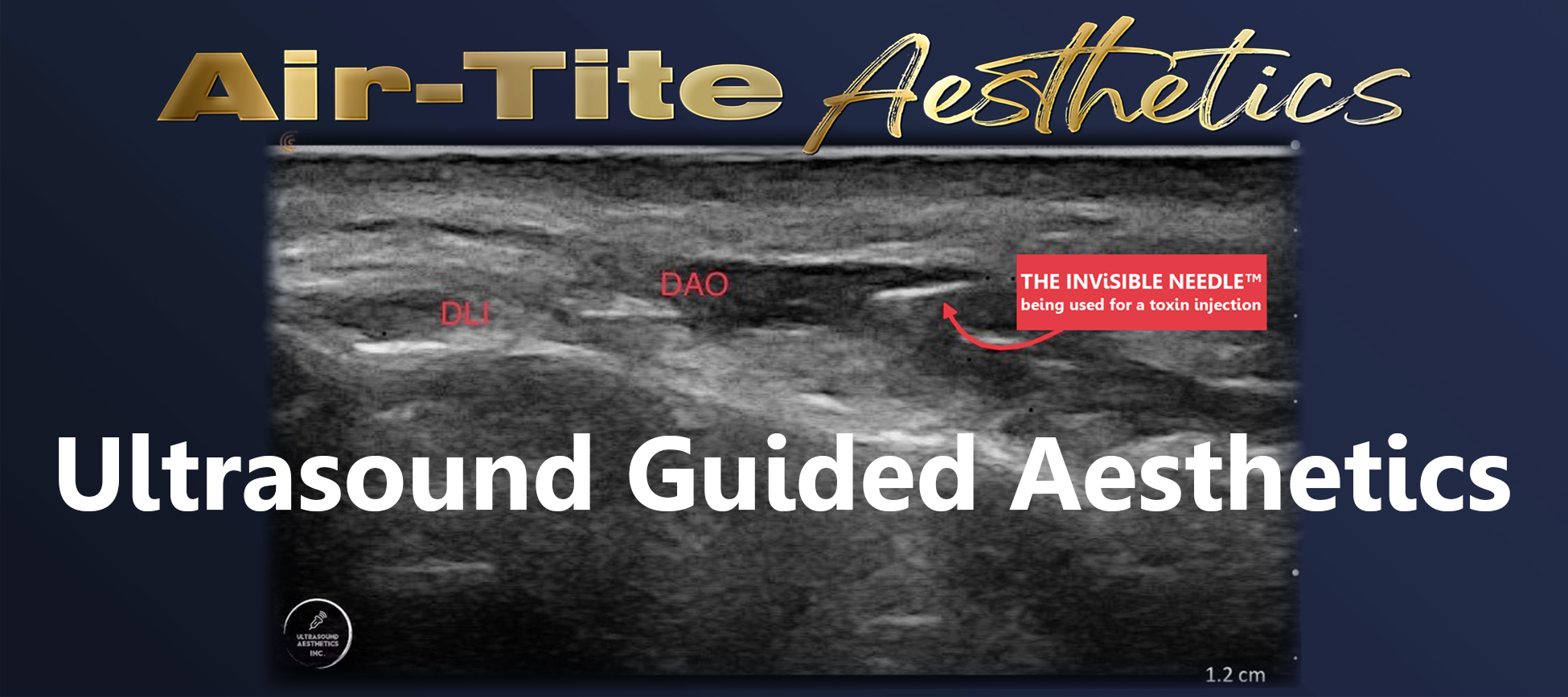
One study from The Aesthetic Surgery Journal discussed this as an optimal method for neurotoxin injections for correcting asymmetrical smiles with the assistance of ultrasound to see the depressor anguli oris (DAO), depressor labii inferioris (DLI), and zygomaticus major (Zmi). Ultrasound for facial aesthetics enables you to clearly see morphology, location, layer of target muscles and thickness of subcutaneous fat tissue allows for the highest accuracy and proper dosing.
A common treatment for TMJ (Temporal Mandibular Joint) disorder & bruxism (excessive teeth grinding) involves injecting neuromodulators into the masseter muscles.
Dr. Arora has found that some patients who previously received masseter injections without ultrasound guidance still experience troublesome jaw pain. Upon ultrasound examination of the masseter muscles, these patients had multiple septa arising from the intermediate tendon. This essentially divided the muscle into different compartments, causing portions of their masseter muscle to be more hyperactive than other areas.
The septa and tendon possibly prevented the spread of the neuromodulator uniformly through the muscle during their unsuccessful previous treatments. Dr. Arora addresses this issue by injecting more units of neurotoxin into the more contractile areas under ultrasound guidance for optimal results. This ultimately provides complete relief to the patient and saves them money, since less units are required overall.
An aesthetic ultrasound exam is a convenient, fast, and non-invasive technique that can greatly benefit your aesthetic practice by improving the assessment and prevention of dermal filler complications. Air-Tite carries a wide variety of products that are essential to help visualize injection placement under ultrasound.
TSK STERiGLIDE™ cannulas are made with a proprietary surface coating that allows greater visibility under sonography. Because the STERiGLIDE™ cannulas also have the nearest-to-tip port delivery, you can confidently inject filler directly into the area you visualize under ultrasound.
Just take it from Dr. Arora who claims "I prefer TSK STERiGLIDE™ cannulas for ultrasound procedures due to two reasons: The first being that TSK cannulas show better on ultrasound, possibly due to the specific coating the cannulas have. Second is that the STERiGLIDE™ cannula port is right next to the tip unlike many other cannulas, and this is a big advantage to give precise injections. Using ultrasound is very rewarding to both the practitioner and patient. The ability to see both the anatomy and the needle during a procedure increases safety and is priceless.”
LEARN MORE ABOUT TSK STERiGLIDE™ CANNULAS