


Your shopping cart is currently empty.
-Written by Erin Ashe, CSA, MSA
Vascular occlusions are dangerous, but the signs that an injection has caused the onset of one are usually present if you know what to look for. Skin coolness, severe pain, and acute onset of skin color changes are generally indicators of a vascular occlusion occurrence.
However, correctly identifying the symptoms is merely the first step in a series of events that must occur to stop the progress of a vascular occlusion. To be prepared to address this critical event if it occurs, you and your team should have protocols in place for how you will handle the problem.
Hyaluronidase will dissolve filler, but too many injectors do not keep it stocked or have a plan in place for using it. Read on to learn more about vascular occlusions and how to quickly address them to reopen the vessel.
Note: This information contained within this blog post is for educational purposes only and does not substitute for professional medical advice.
A Vascular Occlusion is a rare but potentially detrimental complication that occurs during the injection of fillers. It results from accidental injection of filler into an artery or compression of the artery from the surrounding filler material.
This will cause a blockage, stop blood flow, and diffuse necessary tissue oxygen. Vascular occlusion can rapidly develop into tissue necrosis or even more severe complications if not correctly identified—and treated quickly.
In a recent article written in JAMA Dermatology, it was reported that vascular occlusions can happen in any part of the face injected with filler. The most common areas of concern are the glabella, nasal dorsum, forehead, nasolabial folds, and periorbital regions. Skin necrosis is a frequent outcome; however, the most devastating is the injection-related visual compromise, including blindness. While rare, cases of stroke have been reported in the past.
According to the Aesthetic Complications Guidelines written by Dr. Martyn King, et. al in the Journal of Clinical and Aesthetic Dermatology, there are a few initial warning signs that the practitioner or injector should be on the lookout for:
If not treated immediately, the skin will turn dark and dusky, sometimes blueish with fine reticulations or a lace-like appearance to areas that extend farther than the injection site. Vascular occlusions often happen almost immediately during the filler injections, but in some cases, it can take upwards of 12-24 hours.
While the injector should know how to recognize the early onset symptoms of vascular occlusion in the office, the patient should also be asked to report anything unusual as soon as possible. If the provider suspects a vascular occlusion during injection, they should inform the patient that they will need to begin the process to dissolve the filler immediately.
There is a potential to resolve the occlusion in some situations through tapping, massaging, or applying heat to the affected area. However, if those methods fail to work, it’s imperative to administer hyaluronidase immediately.
Implementing treatment more quickly increases the chances that the occlusion resolves before significant damage occurs. It can take a few minutes to several hours to fully dissolve the HA filler with a duration of action lasting 24-48 hours. It may also take several rounds of hyaluronidase injections to dissolve the HA filler completely.
After the initial vascular occlusion protocol steps and injection of hyaluronidase have taken place, and depending on the response of the affected area, the provider may decide to refer the patient to a specialist or administer another round of hyaluronidase injections.
In a recent communication with Dr. Mariale Foley, oculoplastic facial surgeon, we learned in detail what she recommends if vascular compromise associated with vision loss has occurred. First, injection of filler must be immediately discontinued and the patient referred to an Ophthalmology center. In an ideal situation, the patient should be seen by a retina specialist in the first 90 minutes after the event occurred.
The retina is highly sensitive to hypoxia and exposure to it for a longer time causes irreversible ischemia and necrosis. While waiting for the ambulance to transport the patient to the ER, in-office treatments should be performed. They typically involve a quick visual acuity test and pupil examination.
Lowering the intraocular pressure and dislodging the embolus should be attempted by placing the patient in a supine position, and performing an ocular massage. Intraocular lowering pressure agents can be given, such as topical aqueous suppression drops, IV mannitol can also be administered if available. All the treatments applied to the patient need to be notified to the referral Ophthalmology or ER center.
Dr. Foley also states that in case an ophthalmologist is not immediately available and the injector feels comfortable with giving the patient a retrobulbar injection of hyaluronidase, this procedure could be attempted. This technique is explained in the paper, Retro or PeriBulbar Injection Techniques to Reverse Visual Loss After Filler Injections.
Hyaluronic acid fillers are the most common type of dermal filler treatment. But there are other fillers that are non-hyaluronic acid-based and cannot be broken down by Hyaluronidase. An injector must also be mindful of the viscosity and density of the filler they are working with. Fillers with a greater density have a higher likelihood of placing pressure around a vessel.
You can reduce the risk by using smaller increments of the nonreversible product, and lower the viscosity by premixing with lidocaine solution or using a smaller gauge needle.
Fillers such as Sculptra have the principal ingredient poly-L-lactic acid (PLLA), a substance that can boost collagen products in the skin. Other non-hyaluronic acid fillers such as Bellafill are made of polymethylmethacrylate (PMMA). Radiesse is composed of calcium hydroxyapatite (CaHA), and it is also possible to use autologous fat as an HA filler substitute.
There are other protocols to treat a vascular occlusion with a non-HA filler:
Note: You can use all of the above protocols for the hyaluronic acid filler vascular occlusion protocols in conjunction with the hyaluronidase injections.
Dr. Mariale Foley stresses the importance of injectors having a keen understanding of the following to help avoid potential complications:
Having a vascular occlusion strategy checklist is also essential, and the following is made up of essential steps that Dr. Mariale Foley shared with us:
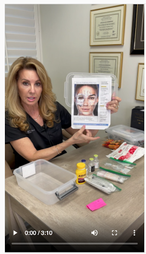
In a recent video, Lori Robertson MSN, FNP-C, and co-founder of The Aesthetic Immersion, shared a video of the vascular occlusion kit that she personally uses in her office.
She highlights the syringes, needles, and the TSK STERiGLIDE™ microcannulas that she gets from Air-Tite and puts into her kit (along with several other items). She mentions that if you use a microcannula to inject the filler where the vascular occlusion occurred, you can also use the microcannula to inject the hyaluronidase.
Lori’s vascular occlusion kit includes:
She has a similar ocular occlusion kit that contains some different items. This kit contains:
At Air-Tite, we sell syringes, microcannulas (STERiGLIDE™ and CSH), and needles that can be used to inject Hyaluronidase, as well as filler. Click below to learn more about our STERiGLIDE™ premium aesthetic cannula.
STERiGLIDE™ premium aesthetic cannulas